
Medicine
Medical imaging
A number of imaging technologies are used to diagnose diseases and guide treatment within our bodies. This branch of medicine is called radiology, and it is practised by medical doctors known as radiologists. It began with the use of X-rays for viewing bones and dense materials, but today includes all types of imaging, including CT scans (the use of X-rays for cross-sectional images), MRI scans (the use of magnets and radio waves to provide images of soft tissue), PET scans (the use a radioactive tracer to reveal how the body is functioning), ultrasound (the use of high-frequency sound waves to create images) and laser Doppler imaging (the use of lasers to create images of live tissue).
X-rays
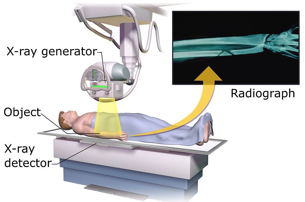
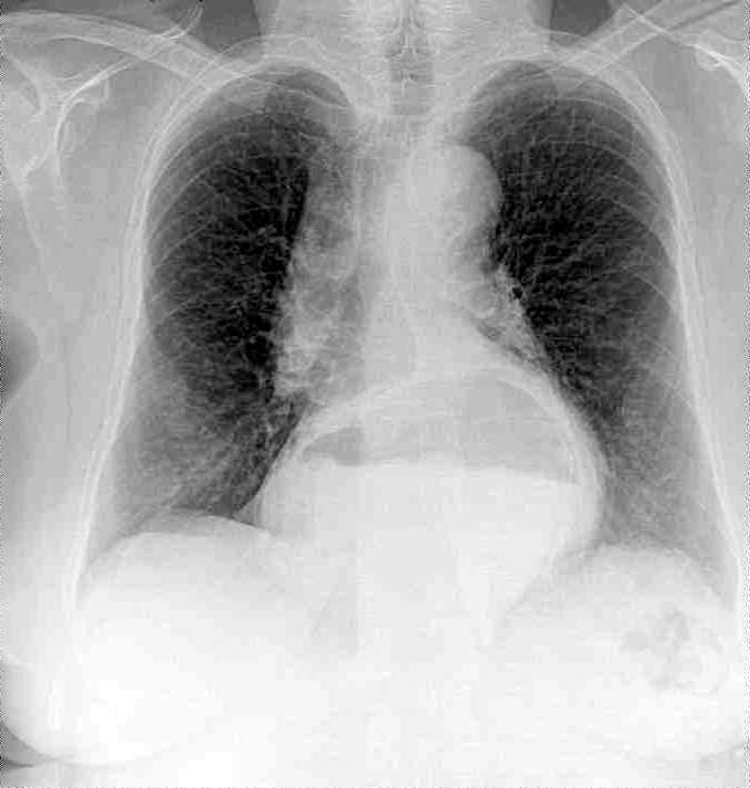
X-ray imaging works by passing a beam of X-rays, a form of electromagnetic radiation, through a part of the body. X-rays have extremely high energy, allowing them to pass right through some solid materials. As the X-rays travel through the body, their energy is absorbed by different parts of the body at different rates. A detector captures the X-rays after they have passed through and turns them into an image. Softer parts of your body that X-rays can pass through easily, such as the heart and lungs, show up as darker areas, while bones, teeth and other dense materials appear white because they block the X-rays.
X-rays are mainly used to look at the teeth, bones and joints, although they are sometimes used to detect problems affecting soft tissue.
CT scans
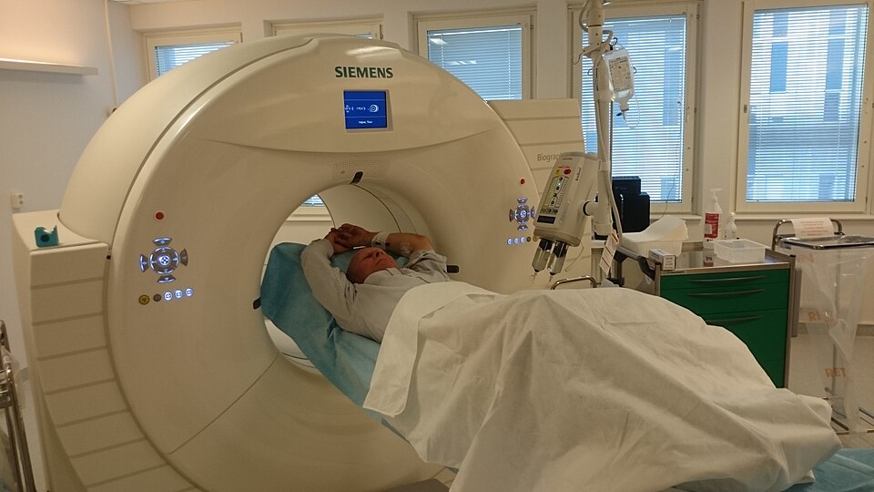
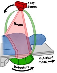
A computerised tomography (CT) scan also uses X-ray beams, but they are rotated around the body. The scanner is a large ring-shaped device, which moves over the patient. Multiple X-rays are captured from different angles. These are then processed on a computer to produce tomographic (cross-section) images, or "slices" of the body. The 3D images contain more information than regular X-rays from a single beam. CT scans can produce detailed images of many structures inside the body, including the internal organs and blood vessels. These may help to determine the location, size and shape of a cancerous tumour, for example, or allow a doctor to take a needle biopsy—where a small tissue sample is removed using a needle.


MRI scans
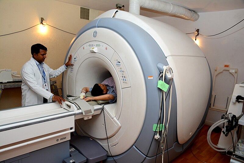
Magnetic resonance imaging (MRI) uses strong magnetic fields and radio waves to produce high-resolution images of the inside of the body, including its soft tissues such as muscles, tendons and blood vessels. Unlike CT, MRI does not use X-rays, but it does provide more detailed images than CT scans. Using MRI scanners, it is possible, for example, to measure how much blood is flowing through the vessels and to detect whether there are any blockages.
Human body tissue is mostly made up of water molecules. These consist of hydrogen and oxygen atoms. At the centre of each hydrogen atom is a subatomic particle, called a proton. Protons are like tiny magnets. In the presence of a magnetic field created by the MRI scanner, they line up in the same direction (similar to the way a needle of a compass is pulled into position by a magnet).
The scanner then emits short bursts of radio waves, which knock the protons out of alignment. When the bursts stop, the protons re-align. Protons re-align at different speeds according to the tissue they are in, and release different signals. Computer software uses this information to pinpoint the exact location of the atoms in the body and to create high-res images of the different tissue types.

PET scans
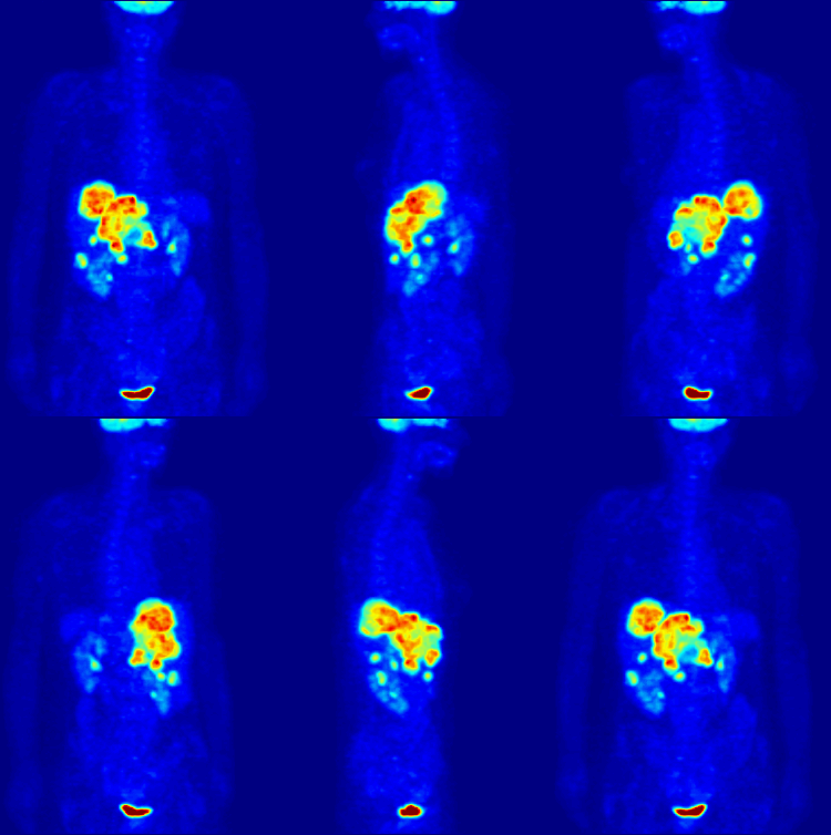
Positron emission tomography (PET) scans are used to produce detailed 3D images of the inside of the body. PET scanners work by detecting the radiation given off by a radioactive tracer, a kind of sugar similar to naturally occurring glucose, as it collects in different parts of the body. Having been injected into the body, the tracer releases radiation that can be measured by a detector.
By analysing the areas where the tracer does (or does not) build up, it is possible to view any abnormalities in the way certain body functions are working. A concentration of tracer in the body’s tissues can help identify cancerous cells. This is because cancer cells use glucose at a much faster rate than normal cells. Sometimes PET scans are used to help plan operations, such as brain surgery. They can also help diagnose some conditions that affect the normal workings of the brain, such as dementia.
Ultrasound
An ultrasound scan, sometimes called a sonogram, is a procedure that uses high-frequency sound waves to create an image of part of the inside of the body. Completely safe since it uses no radiation, ultrasound is often used during pregnancy to monitor the unborn baby. It also used to look at the heart and other organs.


A small device called an ultrasound probe, which gives off high-frequency sound waves, is moved over the skin. (A lubricating gel on the skin allows the probe to move smoothly and ensures there is continuous contact between the probe and the skin.) As the waves from the probe travel though the body, they bounce off different parts. These “echoes” are picked up by the probe and turned into a moving image, which is displayed on a monitor while the scan is carried out.
With an endoscopic ultrasound scan, the probe is attached to a long, thin, flexible tube (an endoscope) which is inserted into the body, usually through the mouth, to examine the oesophagus or stomach. The endoscope has a light and an ultrasound device on the end.


Laser Doppler imaging
Laser Doppler imaging (LDI) uses a laser beam to create images showing the flow of blood through organ tissue. When a laser is shone on to it, the light is scattered by moving red blood cells. The laser light is slightly shifted in frequency (this shift known as the Doppler effect) by the moving cells. This light is detected using a sensor, which converts it into an electrical signal. The strength of the signal reflects the speed and concentration of blood flow. A map can then be created from this information.
LDI is widely used in medicine, including ophthalmology (the diagnosis and treatment of eye diseases and disorders) where it is used to measure blood flow in the retina.