
Human body

CITE
We have made every effort to follow citation style rules, but there may be some minor differences. If in doubt, please refer to the appropriate citation style manual.
Eye. (2026). In Q-files Encyclopedia, Life, Human body. Retrieved from
https://www.q-files.com/life/human-body/eye
"Eye." Life, Human body, Q-files Encyclopedia, 23 Mar. 2026.
https://www.q-files.com/life/human-body/eye.
Accessed 22 Sep. 2026.
Eye 2026. Life, Human body. Retrieved 22 September 2026, from
https://www.q-files.com/life/human-body/eye
Life, Human body, s.v. "Eye," accessed September 22, 2026.
https://www.q-files.com/life/human-body/eye


Eye
Sight is the most important of our senses: around three-quarters of all information processed in the brain comes in through the eyes alone. Each eyeball is about 25 millimetres (1 inch) across and well protected in a socket, called the orbit, inside the skull bone. Light rays enter the eye through its transparent domed front, the cornea. They pass through the pupil, a hole in a ring of muscle known as the iris. The rays are bent or focused by the lens, and shine a clear image on to the retina which lines the rear of the eyeball. When light hits the retina, light-sensitive cells produce nerve signals, which pass along the optic nerve to the brain.

How the eye works
Light enters the eye through a transparent, domed window called the cornea at the front of the eye. This does most of the work of focusing images on to the retina. Only half a millimetre thick at the centre, its outer surface is the epithelium, a transparent continuation of the skin. The cornea thickens to one millimetre where it joins the white of the eye, called the sclera.
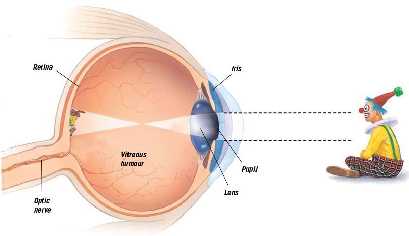

Light passes through an opening, called the pupil, in the iris, the eye’s coloured part. This is a ring of muscle that controls the size of the pupil. The iris works like the aperture on a camera: if too much light enters the eye, the pupil is reduced in size. If too little light enters, it grows larger. The pupil can vary in size from one to eight millimetres (0.04–0.3 inches). The adjustment is automatic—it works without any conscious effort.
Behind the iris there is a soft, elastic lens, which finely adjusts the sharpness of the image. The main body of the eye is filled with a clear, jelly-like substance called the vitreous humour. This gives the eye its shape and firmness. Light shines through this on to the retina.
Retina
The retina is the thin layer that lines the back of the eyeball. It contains light-sensitive cells—about 130 million of them—that send signals back along the optic nerve, a bundle of about a million nerve cells, to the brain. The fovea, a region in the centre of the retina directly behind the lens, is the place where the eye best focuses images from the outside world. Because the light rays cross over in the eye, the image is upside down. The brain tells us how the world really looks.

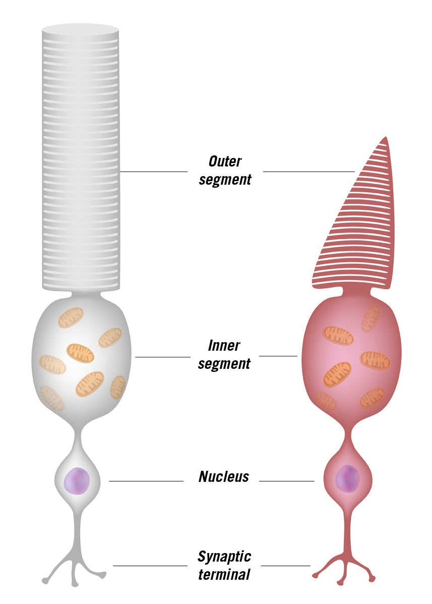
The retina is made up of two different types of light-sensitive cells, called rods and cones after their respective shapes. Rods are more sensitive to intensity of light than to colour, which is the job of the cones. Cones are most abundant at the fovea, the part of the retina where the clearest, most colourful images are created. There are no rods and cones at the point where the nerves leave the eye to form the optic nerve. This area of the retina is called the optic disc, or blind spot. It is also the place where the blood vessels that supply the retina enter the eye.
Moving the eye
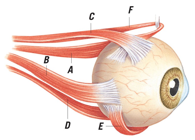
Six small, strap-shaped muscles attach the eyeball to the rear of the eye socket. They work together to move the eyeball in different directions. Muscle A swivels it to the left, B to the right, C upwards and D downwards. Muscles E and F rotate the eyeball diagonally.
Consultant: Richard Walker
pics
Without in any way limiting Q-files Ltd’s exclusive rights under copyright, any use of this publication to “train” generative artificial intelligence (AI) technologies to generate text is expressly prohibited. Q-files Ltd reserves all rights to license use of this work for generative AI training and development of machine learning language models.